Breast Cancer

Mammogram

In a mammogram, the patient is directed to stand in front of an x-ray machine. Your breast is placed between two plastic plates that flatten your breast to get a clear image.

(This procedure is known to be slightly discomforting)

2D mammograms - Takes standard X-ray images from two angles per breast, producing one flat image.

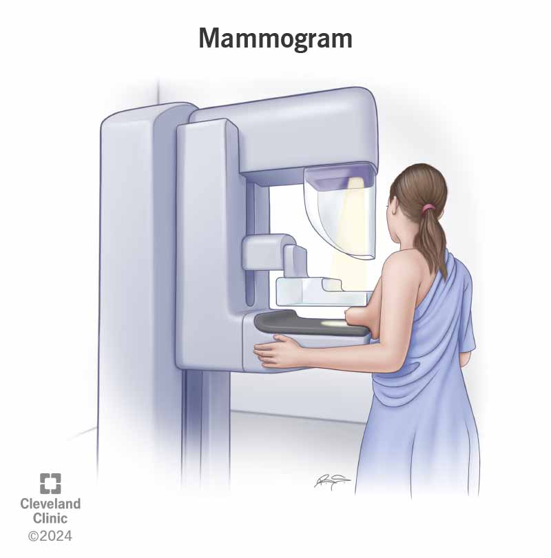
This is the most common screening method for breast cancer

3D mammograms/Digital Breast Tomosynthesis (DBT) - An X-ray tube moves in an arc, capturing multiple low-dose images from various angles

This option has improved detection and fewer false alarms!

Screening Frequency (average-risk patient*):

*Average risk patient
  • No personal history of breast cancer
  • No known genetic mutation (like BRCA1 or BRCA2)
  • No strong family history of breast, ovarian, pancreatic, or prostate cancer
  • No chest radiation at a young age
  • Screening should begin at age 40 (at most) for all women
  • Ages 40-44 → Annual or biannual, depends on test results at age 40
  • Ages 45-54 → Annual
  • Ages 55+ → Every 2 years

For high-risk patients*:

**High-risk patient
  • BRCA1/2 mutation or other high-risk genes
  • Strong family history of breast/related cancers
  • Chest radiation at a young age
  • Start at age 30
  • Recommended to get both a breast MRI and mammogram annually

Breast MRI (magnetic resonance imaging)

The MRI machine makes detailed 3D images of your breast when you lay down (on your belly) on a table and are imaged inside an MRI machine.

(This procedure may require fluids injected through in IV, which can help make better images of the inside of the breast → brief pain)

Breast MRI procedure illustration

Source: Cleveland Clinic

Screening Frequency (average risk patient):

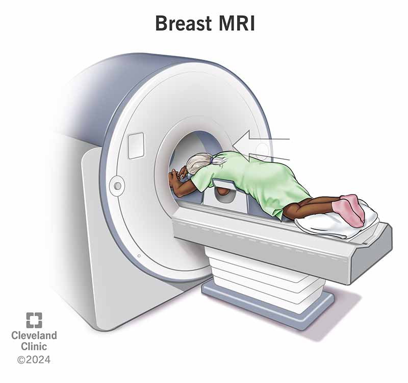
A breast MRI is not done for average risk patients, only mammograms!

Screening Frequency (high risk patient):

  • → A Breast MRI is not recommended as a screening test by itself, it should be paired with a mammogram in order to be most effective
  • Start at age 30 and do MRI along with a mammogram

Clinical Breast Exam

A doctor will physically examine breasts to feel for any abnormal spots.

(Women should be familiar with how their breasts normally look and feel and should report any changes to a health care provider right away)

Clinical breast exam illustration

Source: wikiHow

***This screening method doesn't have strong effectiveness for average risk patients and is typically paired with a mammogram to get clearer imaging (not a replacement for mammograms and breast MRIs)

Screening Frequency (average risk patient):

  • Does not need to be done annually, no routine checkups
  • Not recommended at any age but is utilized when the patient has a suspicion/notices lumps or abnormalities in breast appearance

Screening Frequency (high risk patient):

  • Usually done every 6-12 months starting at age 30
  • Individualized routine, discuss with your doctor

More Resources: